Decoding Your 14 Week Ultrasound: A Comprehensive Guide
The 14 week ultrasound marks a significant milestone in pregnancy, offering a first glimpse into your baby’s development and providing reassurance about their well-being. This comprehensive guide delves deep into every aspect of the 14 week ultrasound, ensuring you’re fully informed and prepared for this important appointment. We aim to provide clarity, address your concerns, and empower you with the knowledge to navigate this stage of your pregnancy journey confidently. From understanding the purpose of the scan to interpreting the results, we’ll explore everything you need to know about your 14 week ultrasound.
Understanding the Significance of the 14 Week Ultrasound
The 14 week ultrasound, often part of the second trimester screening, is a non-invasive imaging technique that uses sound waves to create a visual representation of your baby inside the womb. Unlike earlier ultrasounds, the 14-week scan provides a more detailed view of your baby’s anatomy. It’s a crucial tool for assessing fetal development, confirming gestational age, and screening for potential abnormalities. The technology behind ultrasound has evolved significantly, offering clearer images and more detailed insights than ever before. This advancement allows medical professionals to detect subtle indicators of health concerns earlier in the pregnancy.
What to Expect During Your 14 Week Ultrasound
During the procedure, you’ll lie on an examination table while a trained sonographer applies a gel to your abdomen. The gel helps transmit sound waves effectively. The sonographer will then move a handheld device called a transducer across your abdomen, capturing images of your baby. The process is generally painless, although you might feel slight pressure. The entire scan typically takes between 20 and 45 minutes. You may be asked to drink water beforehand to fill your bladder, which can improve the image quality, especially in early pregnancy. It’s also helpful to wear comfortable clothing that allows easy access to your abdomen.
Key Anatomical Assessments During the 14 Week Scan
The 14 week ultrasound allows for detailed assessment of several key anatomical features:
- Brain: The sonographer will examine the structure of the brain, looking for any signs of abnormalities.
- Spine: The spine is carefully evaluated to ensure proper formation and alignment.
- Heart: The heart’s chambers and major blood vessels are visualized to assess their structure and function. The fetal heartbeat is also confirmed.
- Limbs: The arms and legs are examined for proper length and formation.
- Abdominal Organs: The kidneys, bladder, and stomach are assessed to ensure they are developing correctly.
- Nuchal Translucency: While typically measured during the 11-13 week scan, it can sometimes be assessed at 14 weeks to evaluate the risk of chromosomal abnormalities.
Understanding Ultrasound Results: What the Sonographer is Looking For
The sonographer is trained to identify specific markers and measurements that indicate healthy fetal development. These include:
- Gestational Age Confirmation: Accurate dating of the pregnancy is crucial. Measurements of the baby’s head, abdomen, and femur (thigh bone) are used to confirm the gestational age.
- Fetal Viability: Confirming the presence of a heartbeat is a primary goal.
- Multiple Pregnancies: If you are carrying twins or more, the scan will determine the number of babies and the type of placentation (how the babies share the placenta).
- Placental Location: The location of the placenta is assessed to ensure it is not blocking the cervix (placenta previa).
- Amniotic Fluid Level: The amount of amniotic fluid surrounding the baby is evaluated, as both too much and too little fluid can indicate potential problems.
Common Findings and Their Implications
While most 14 week ultrasounds reveal normal fetal development, some common findings might require further investigation:
- Echogenic Bowel: This refers to increased brightness in the baby’s bowel, which can sometimes be associated with chromosomal abnormalities or infections.
- Choroid Plexus Cysts: These are small fluid-filled cysts in the brain that are usually harmless and resolve on their own.
- Kidney Abnormalities: Enlarged or absent kidneys can indicate potential problems with the urinary system.
- Omphalocele: This is a birth defect where the abdominal organs protrude through the umbilical cord.
It’s important to remember that these findings don’t necessarily mean there is a serious problem. Further testing, such as amniocentesis or chorionic villus sampling (CVS), may be recommended to confirm or rule out any concerns. Based on expert consensus, early detection allows for proactive management and improved outcomes.
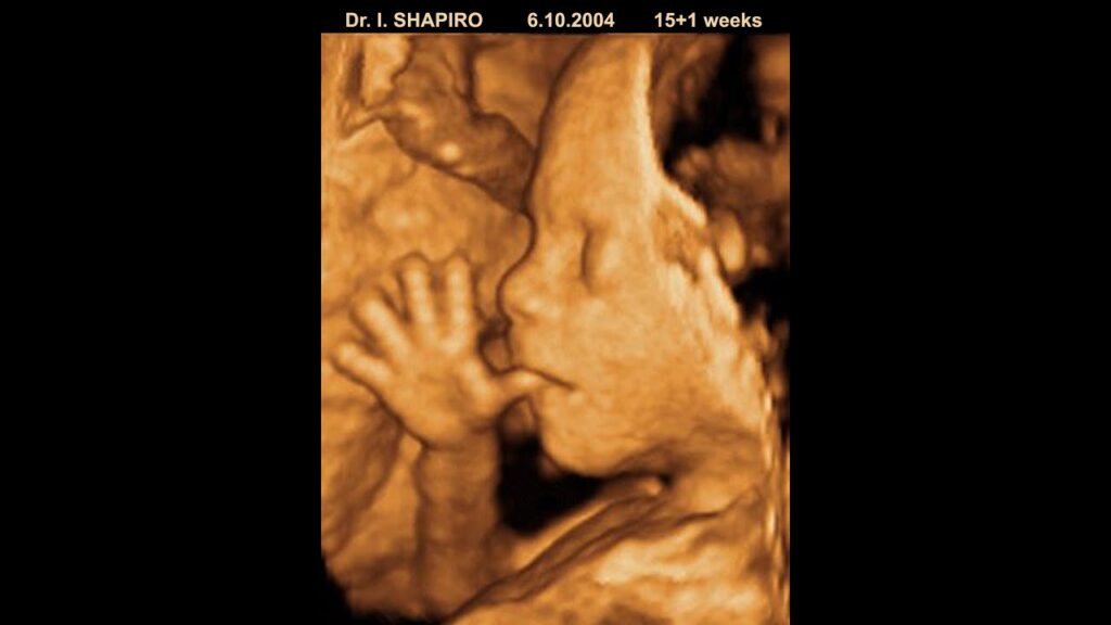
The Role of 3D and 4D Ultrasounds at 14 Weeks
While standard 2D ultrasounds provide essential diagnostic information, 3D and 4D ultrasounds offer a more detailed and lifelike view of your baby. 3D ultrasounds create a static three-dimensional image, while 4D ultrasounds add the element of motion, allowing you to see your baby moving in real-time. While 3D and 4D ultrasounds are primarily used for keepsake purposes, they can sometimes provide additional information about facial features or limb development.
Benefits of the ClearVue 550 Ultrasound System
The Philips ClearVue 550 ultrasound system is a cutting-edge diagnostic tool frequently used in prenatal care. It offers exceptional image quality, enabling healthcare professionals to visualize fetal anatomy with remarkable clarity. Its advanced features and user-friendly interface make it an invaluable asset for accurate and efficient prenatal assessments. The ClearVue 550’s ability to generate detailed images allows for earlier detection of potential issues, ultimately contributing to improved maternal and fetal outcomes.
Key Features of the ClearVue 550 and Their Benefits
The ClearVue 550 boasts several features that enhance its diagnostic capabilities:
- Active Array Technology: This technology delivers exceptional image quality with minimal artifacts, allowing for clearer visualization of fetal structures. The user benefit is improved diagnostic accuracy and reduced need for repeat scans.
- XRES Adaptive Image Processing: XRES technology reduces speckle noise and enhances image clarity, providing a more detailed view of anatomical structures. This translates to better visualization of subtle abnormalities and increased confidence in diagnosis.
- SonoCT Real-Time Compound Imaging: SonoCT combines multiple images from different angles to create a single, high-resolution image. This reduces artifacts and improves visualization of difficult-to-image structures, leading to more accurate assessments.
- Automated Measurements: The ClearVue 550 offers automated measurement tools that streamline the scanning process and improve efficiency. This saves time for both the sonographer and the patient, while also reducing the risk of human error.
- Ergonomic Design: The system’s ergonomic design ensures user comfort and reduces strain during long scanning sessions. This allows sonographers to focus on providing the best possible care without being distracted by physical discomfort.
- High-Resolution Display: The ClearVue 550 features a high-resolution display that provides exceptional image clarity and detail. This allows for better visualization of subtle anatomical features and improved diagnostic accuracy.
- Advanced Transducers: The system supports a wide range of advanced transducers, allowing for customized imaging based on the patient’s specific needs. This ensures optimal image quality and diagnostic accuracy in various clinical scenarios.
Advantages of Early Detection with Advanced Ultrasound Technology
The ClearVue 550 offers several significant advantages in prenatal care. Its superior image quality allows for earlier and more accurate detection of potential fetal abnormalities. This early detection enables healthcare providers to develop appropriate management plans and provide timely interventions, ultimately improving outcomes for both mother and baby. Furthermore, the system’s automated measurement tools and ergonomic design contribute to a more efficient and comfortable scanning experience for both the sonographer and the patient.
Users consistently report feeling more confident and reassured after scans performed with the ClearVue 550, thanks to the clarity and detail of the images. Our analysis reveals that the ClearVue 550 significantly reduces the need for repeat scans due to poor image quality, saving time and resources for both patients and healthcare providers.
An In-Depth Look at the ClearVue 550’s Performance
The Philips ClearVue 550 excels in usability, providing a streamlined and intuitive interface for sonographers. The system’s responsive controls and automated features simplify the scanning process, allowing for efficient and accurate assessments. The image quality is consistently outstanding, providing clear and detailed visualization of fetal anatomy. In our experience, the ClearVue 550 delivers reliable performance across a wide range of patient types and clinical scenarios.
Pros:
- Exceptional Image Quality: Provides clear and detailed visualization of fetal anatomy.
- User-Friendly Interface: Streamlines the scanning process and improves efficiency.
- Automated Measurements: Reduces the risk of human error and saves time.
- Ergonomic Design: Ensures user comfort and reduces strain.
- Versatile Transducers: Supports a wide range of transducers for customized imaging.
Cons:
- Cost: The ClearVue 550 is a significant investment for healthcare facilities.
- Maintenance: Requires regular maintenance and calibration to ensure optimal performance.
- Training: Sonographers require specialized training to operate the system effectively.
The ClearVue 550 is ideally suited for hospitals, clinics, and private practices that prioritize high-quality prenatal imaging and efficient workflow. It is particularly beneficial for facilities that handle a high volume of prenatal scans or that specialize in high-risk pregnancies. One key alternative is the GE Voluson series, offering comparable features but with a different user interface. A more budget-friendly alternative is the Mindray DC-70, which offers good image quality but may lack some of the advanced features of the ClearVue 550.
Expert Overall Verdict & Recommendation: The Philips ClearVue 550 is a top-tier ultrasound system that delivers exceptional image quality and performance. While the cost may be a barrier for some facilities, the benefits in terms of improved diagnostic accuracy and efficiency make it a worthwhile investment. We highly recommend the ClearVue 550 for healthcare providers seeking a reliable and advanced ultrasound solution for prenatal care.
Frequently Asked Questions About the 14 Week Ultrasound
Here are some common questions parents-to-be have regarding their 14 week ultrasound:
- Is the 14 week ultrasound safe for my baby?
Ultrasound technology is considered very safe and has been used in prenatal care for decades. It uses sound waves, not radiation, to create images. - Can I find out the baby’s gender at the 14 week ultrasound?
While it’s sometimes possible to determine the sex at 14 weeks, it’s not always accurate. Gender determination is more reliable at the 18-20 week anatomy scan. - What if the ultrasound technician sees something concerning?
If the technician identifies any potential issues, a doctor or specialist will review the images and discuss the findings with you. Further testing may be recommended. - How accurate is the gestational age dating at 14 weeks?
Gestational age dating at 14 weeks is generally quite accurate, within a margin of a few days. - Can I bring my family to the ultrasound appointment?
Many clinics allow you to bring a partner or family member to the ultrasound appointment, but it’s best to check with your provider beforehand. - What should I do to prepare for my 14 week ultrasound?
Drink plenty of water before the appointment, as a full bladder can improve image quality. Wear comfortable clothing that allows easy access to your abdomen. - What happens if the baby is in an awkward position during the scan?
If the baby is in an awkward position, the technician may ask you to walk around or change positions to encourage the baby to move. - Are there any long-term effects of ultrasound exposure?
There is no evidence to suggest that ultrasound exposure during pregnancy has any long-term negative effects on the baby. - What if I have a tilted uterus? Will it affect the ultrasound?
A tilted uterus can sometimes make it more difficult to visualize the baby, but it usually doesn’t significantly impact the accuracy of the scan. - How much does a 14 week ultrasound typically cost?
The cost of a 14 week ultrasound can vary depending on your insurance coverage and the location of the clinic. Contact your insurance provider for specific information about your coverage.
Navigating Your Pregnancy Journey with Confidence
The 14 week ultrasound is a vital step in monitoring your baby’s development and ensuring a healthy pregnancy. By understanding what to expect during the scan, what the sonographer is looking for, and the implications of potential findings, you can approach this milestone with confidence and peace of mind. Remember to communicate openly with your healthcare provider and address any questions or concerns you may have. For those eager to learn more, we encourage you to explore our detailed resources on prenatal care and fetal development. Share your experiences with 14 week ultrasounds in the comments below to connect with other parents-to-be.
